
The Structure of the Nervous System
The mammalian nervous system is divided into central and peripheral nervous systems.
The Peripheral Nervous System
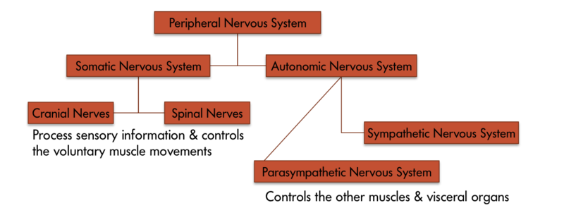
Figure 3 The various components of the peripheral nervous system
The peripheral nervous system is divided into somatic and autonomic nervous systems (Figure 3). Where the somatic nervous system consists of cranial nerves (12 pairs) and spinal nerves (31 pairs) and is under the volitional control of the individual in maneuvering bodily muscles, the autonomic nervous system also running through these nerves lets the individual have little control over muscles and glands. Main divisions of the autonomic nervous system that control visceral structures are the sympathetic and parasympathetic nervous systems.
At an appropriate cue (say a fear-inducing object like a snake), the sympathetic division generally energizes many muscles (e.g., heart) and glands (e.g., adrenals), causing activity and release of hormones that lead the individual to negotiate the fear-causing snake with fight- or-flight responses. Whether the individual decides to fight the snake or run away from it, either action requires energy; in short, the sympathetic nervous system says “go, go, go.” The parasympathetic nervous system, on the other hand, curtails undue energy mobilization into muscles and glands and modulates the response by saying “stop, stop, stop.” This push–pull tandem system regulates fight-or-flight responses in all of us.
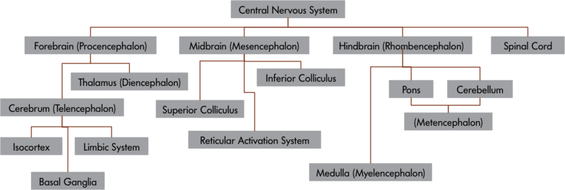
Figure 4 the central nervous system and its components
The Central Nervous System
The central nervous system is divided into a number of important parts (see Figure 4), including the spinal cord, each specialized to perform a set of specific functions. Telencephalon or cerebrum is a newer development in the evolution of the mammalian nervous system. In humans, it is about the size of a large napkin and when crumpled into the skull, it forms furrows called sulci (singular form, sulcus). The bulges between sulci are called gyri (singular form, gyrus). The cortex is divided into two hemispheres, and each hemisphere is further divided into four lobes (Figure 5a), which have specific functions. The division of these lobes is based on two delineating sulci: the central sulcus divides the hemisphere into frontal and parietal-occipital lobes and the lateral sulcus marks the temporal lobe, which lies below.
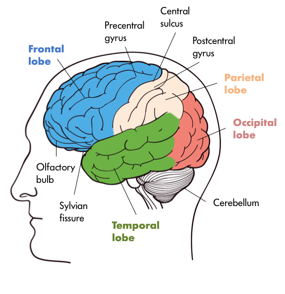
Figure 5a The lobes of the brain
Just in front of the central sulcus lies an area called the primary motor cortex (precentral gyrus), which connects to the muscles of the body, and on volitional command moves them. From mastication to movements in the genitalia, the body map is represented on this strip (Figure 5b).
Some body parts, like fingers, thumbs, and lips, occupy a greater representation on the strip than, say, the trunk. This disproportionate representation of the body on the primary motor cortex is called the magnification factor (Rolls & Cowey, 1970) and is seen in other motor and sensory areas. At the lower end of the central sulcus, close to the lateral sulcus, lies the Broca’s area (Figure 6b) in the left frontal lobe, which is involved with language production. Damage to this part of the brain led Pierre Paul Broca, a French neuroscientist in 1861, to document many different forms of aphasias, in which his patients would lose the ability to speak or would retain partial speech impoverished in syntax and grammar (AAAS, 1880). It is no wonder that others have found subvocal rehearsal and central executive processes of working memory in this frontal lobe (Smith & Jonides, 1997, 1999).
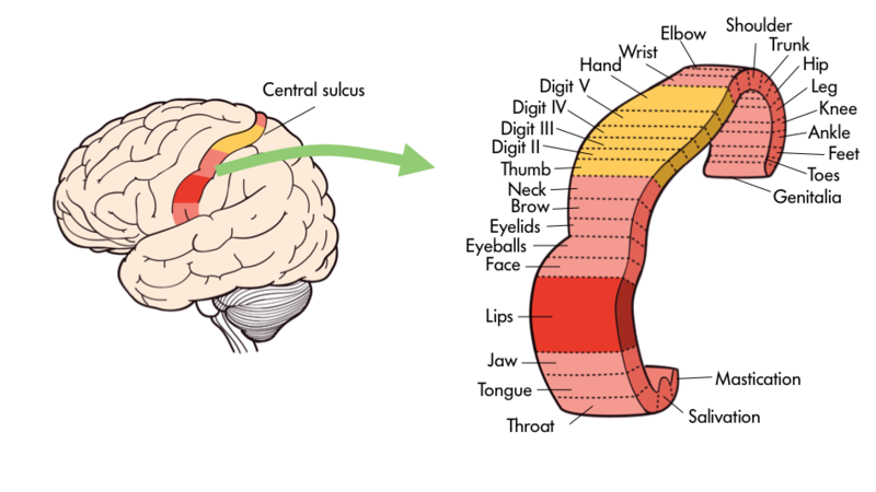
Figure 5b. Specific body parts like the tongue or fingers are mapped onto certain areas of the brain including the primary motor cortex.
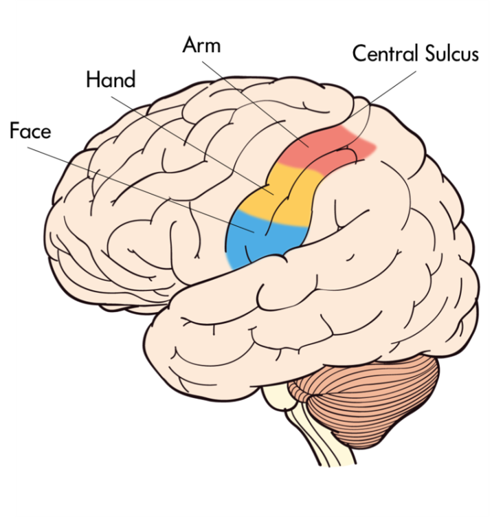
Figure 6a The Primary Somatosensory Cortex
Just behind the central gyrus, in the parietal lobe, lies the primary somatosensory cortex (Figure 6a) on the postcentral gyrus, which represents the whole body receiving inputs from the skin and muscles. The primary somatosensory cortex parallels, abuts, and connects heavily to the primary motor cortex and resembles it in terms of areas devoted to bodily representation. All spinal and some cranial nerves (e.g., the facial nerve) send sensory signals from skin (e.g., touch) and muscles to the primary somatosensory cortex. Close to the lower (ventral) end of this strip, curved inside the parietal lobe, is the taste area (secondary somatosensory cortex), which is involved with taste experiences that originate from the tongue, pharynx, epiglottis, and so forth.
Just below the parietal lobe, and under the caudal end of the lateral fissure, in the temporal lobe, lies the Wernicke’s area (Demonet et al., 1992). This area is involved with language comprehension and is connected to the Broca’s area through the arcuate fasciculus, nerve fibers that connect these two regions. Damage to the Wernicke’s area (Figure 6b) results in many kinds of agnosias; agnosia is defined as an inability to know or understand language and speech-related behaviors. So an individual may show word deafness, which is an inability to recognize spoken language, or word blindness, which is an inability to recognize written or printed language. Close in proximity to the Wernicke’s area is the primary auditory cortex, which is involved with audition, and finally the brain region devoted to smell (olfaction) is tucked away inside the primary olfactory cortex (prepyriform cortex).
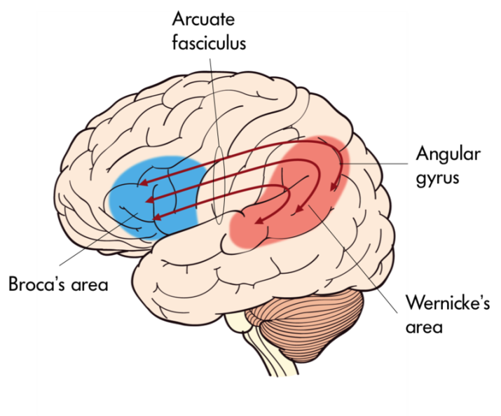
Figure 6b Wernicke’s area
At the very back of the cerebral cortex lies the occipital lobe housing the primary visual cortex. Optic nerves travel all the way to the thalamus (lateral geniculate nucleus, LGN) and then to visual cortex, where images that are received on the retina are projected (Hubel, 1995).
In the past 50 to 60 years, visual sense and visual pathways have been studied extensively, and our understanding about them has increased manifold. We now understand that all objects that form images on the retina are transformed (transduction) in neural language handed down to the visual cortex for further processing. In the visual cortex, all attributes (features) of the image, such as the color, texture, and orientation, are decomposed and processed by different visual cortical modules (Van Essen, Anderson & Felleman, 1992) and then recombined to give rise to singular perception of the image in question.
If we cut the cerebral hemispheres in the middle, a new set of structures come into view. Many of these perform different functions vital to our being. For example, the limbic system contains a number of nuclei that process memory (hippocampus and fornix) and attention and emotions (cingulate gyrus); the globus pallidus is involved with motor movements and their coordination; the hypothalamus and thalamus are involved with drives, motivations, and trafficking of sensory and motor throughputs. The hypothalamus plays a key role in regulating endocrine hormones in conjunction with the pituitary gland that extends from the hypothalamus through a stalk (infundibulum).
As we descend down the thalamus, the midbrain comes into view with superior and inferior colliculi, which process visual and auditory information, as does the substantia nigra, which is involved with notorious Parkinson’s disease, and the reticular formation regulating arousal, sleep, and temperature. A little lower, the hindbrain with the pons processes sensory and motor information employing the cranial nerves, works as a bridge that connects the cerebral cortex with the medulla, and reciprocally transfers information back and forth between the brain and the spinal cord.
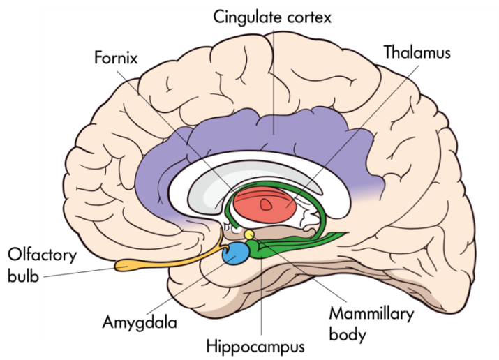
Figure 7 The interior of the brain
The medulla oblongata processes breathing, digestion, heart and blood vessel function, swallowing, and sneezing. The cerebellum controls motor movement coordination, balance, equilibrium, and muscle tone.
The midbrain and the hindbrain, which make up the brain stem, culminate in the spinal cord. Whereas inside the cerebral cortex, the gray matter (neuronal cell bodies) lies outside and white matter (myelinated axons) inside; in the spinal cord this arrangement reverses, as the gray matter resides inside and the white matter outside. Paired nerves (ganglia) exit the spinal cord, some closer in direction towards the back (dorsal) and others towards the front (ventral). The dorsal nerves (afferent) receive sensory information from skin and muscles, and ventral nerves (efferent) send signals to muscles and organs to respond.
