
Vision
How vision works
Vision is a tricky matter. When we see a pizza, a feather, or a hammer, we are actually seeing light bounce off that object and into our eye. Light enters the eye through the pupil, a tiny opening behind the cornea. The pupil regulates the amount of light entering the eye by contracting (getting smaller) in bright light and dilating (getting larger) in dimmer light. Once past the pupil, light passes through the lens, which focuses an image on a thin layer of cells in the back of the eye, called the retina.
Because we have two eyes in different locations, the image focused on each retina is from a slightly different angle (binocular disparity), providing us with our perception of 3D space (binocular vision). You can appreciate this by holding a pen in your hand, extending your arm in front of your face, and looking at the pen while closing each eye in turn. Pay attention to the apparent position of the pen relative to objects in the background. Depending on which eye is open, the pen appears to jump back and forth! This is how video game manufacturers create the perception of 3D without special glasses; two slightly different images are presented on top of one another.
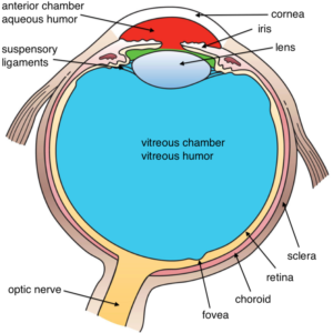
It is in the retina that light is transduced, or converted into electrical signals, by specialized cells called photoreceptors. The retina contains two main kinds of photoreceptors: rodsand cones. Rods are primarily responsible for our ability to see in dim light conditions, such as during the night. Cones, on the other hand, provide us with the ability to see color and fine detail when the light is brighter. Rods and cones differ in their distribution across the retina, with the highest concentration of cones found in the fovea (the central region of focus), and rods dominating the periphery (see Figure 2). The difference in distribution can explain why looking directly at a dim star in the sky makes it seem to disappear; there aren’t enough rods to process the dim light!Next, the electrical signal is sent through a layer of cells in the retina, eventually traveling down the optic nerve. After passing through the thalamus, this signal makes it to the primary visual cortex, where information about light orientation and movement begin to come together (Hubel & Wiesel, 1962). Information is then sent to a variety of different areas of the cortex for more complex processing. Some of these cortical regions are fairly specialized—for example, for processing faces (fusiform face area) and body parts (extrastriate body area). Damage to these areas of the cortex can potentially result in a specific kind of agnosia, whereby a person loses the ability to perceive visual stimuli. A great example of this is illustrated in the writing of famous neurologist Dr. Oliver Sacks; he experienced prosopagnosia, the inability to recognize faces. These specialized regions for visual recognition comprise the ventral pathway (also called the “what” pathway). Other areas involved in processing location and movement make up the dorsal pathway (also called the “where” pathway). Together, these pathways process a large amount of information about visual stimuli (Goodale & Milner, 1992). Phenomena we often refer to as optical illusions provide misleading information to these “higher” areas of visual processing (see Additional Resources for websites containing amazing optical illusions).
Dark and light adaptation
Humans have the ability to adapt to changes in light conditions. As mentioned before, rods are primarily involved in our ability to see in dim light. They are the photoreceptors responsible for allowing us to see in a dark room. You might notice that this night vision ability takes around 10 minutes to turn on, a process called darkadaptation. This is because our rods become bleached in normal light conditions and require time to recover. We experience the opposite effect when we leave a dark movie theatre and head out into the afternoon sun. During lightadaptation, a large number of rods and cones are bleached at once, causing us to be blinded for a few seconds. Light adaptation happens almost instantly compared with dark adaptation. Interestingly, some people think pirates wore a patch over one eye in order to keep it adapted to the dark while the other was adapted to the light. If you want to turn on a light without losing your night vision, don’t worry about wearing an eye patch, just use a red light; this wavelength doesn’t bleach your rods.
Color vision

Our cones allow us to see details in normal light conditions, as well as color. We have cones that respond preferentially, not exclusively, for red, green and blue (Svaetichin, 1955). This trichromatic theory is not new; it dates back to the early 19th century (Young, 1802; Von Helmholtz, 1867). This theory, however, does not explain the odd effect that occurs when we look at a white wall after staring at a picture for around 30 seconds. Try this: stare at the image of the flag in Figure 3 for 30 seconds and then immediately look at a sheet of white paper or a wall. According to the trichromatic theory of color vision, you should see white when you do that. Is that what you experienced? As you can see, the trichromatic theory doesn’t explain the afterimage you just witnessed. This is where the opponent-process theory comes in (Hering, 1920). This theory states that our cones send information to retinal ganglion cells that respond to pairs of colors (red-green, blue-yellow, black-white). These specialized cells take information from the cones and compute the difference between the two colors—a process that explains why we cannot see reddish-green or bluish-yellow, as well as why we see afterimages. Color deficient vision can result from issues with the cones or retinal ganglion cells involved in color vision.
