
The Anatomy of the Brain
There are many ways to subdivide the mammalian brain, resulting in some inconsistent and ambiguous nomenclature over the history of neuroanatomy (Swanson, 2000). For simplicity, we will divide the brain into three basic parts: the brain stem, cerebellum, and cerebral hemispheres (see Figure 1). In Figure 2, however, we depict other prominent groupings (Swanson, 2000) of the six major subdivisions of the brain (Kandal, Schwartz, & Jessell, 2000).
Brain Stem
The brain stem is sometimes referred to as the “trunk” of the brain. It is responsible for many of the neural functions that keep us alive, including regulating our respiration (breathing), heart rate, and digestion. In keeping with its function, if a patient sustains severe damage to the brain stem he or she will require “life support” (i.e., machines are used to keep him or her alive). Because of its vital role in survival, in many countries a person who has lost brain stem function is said to be “brain dead,” although other countries require significant tissue loss in the cortex (of the cerebral hemispheres), which is responsible for our conscious experience, for the same diagnosis. The brain stem includes the medulla, pons, midbrain, and diencephalon (which consists of thalamus and hypothalamus). Collectively, these regions also are involved in our sleep–wake cycle, some sensory and motor function, as well as growth and other hormonal behaviors.
Cerebellum
The cerebellum is the distinctive structure at the back of the brain. The Greek philosopher and scientist Aristotle aptly referred to it as the “small brain” (“parencephalon” in Greek, “cerebellum” in Latin) in order to distinguish it from the “large brain” (“encephalon” in Greek, “cerebrum” in Latin). The cerebellum is critical for coordinated movement and posture. More recently, neuroimaging studies (see “Studying the Human Brain”) have implicated it in a range of cognitive abilities, including language.
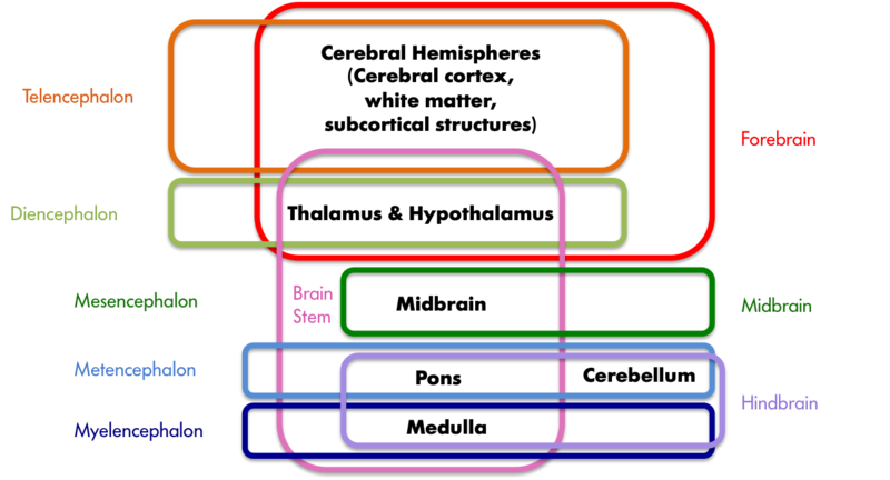
Figure 2. A sample of neuroanatomy nomenclature. The colored boxes indicate the different groupings of the seven structures printed in black, with the labels matching the color of the boxes. The hindbrain, midbrain, and forebrain nomenclature stems from the development of the vertebrate brain; these three areas differentiate early in embryonic development and later give rise to the structures listed in black. These three areas further subdivide into the telencephalon, diencephalon, mesencephalon, metencephalon, and myelencephalon at a later stage of development.
It is perhaps not surprising that the cerebellum’s influence extends beyond that of movement and posture, given that it contains the greatest number of neurons of any structure in the brain. However, the exact role it plays in these higher functions is still a matter of further study.
Cerebral Hemispheres
The cerebral hemispheres are responsible for our cognitive abilities and conscious experience. They consist of the cerebral cortex and accompanying white matter (“cerebrum” in Latin) as well as the subcortical structures of the basal ganglia, amygdala, and hippocampal formation. The cerebral cortex is the largest and most visible part of the brain, retaining the Latin name (cerebrum) for “large brain” that Aristotle coined. It consists of two hemispheres (literally two half spheres) and gives the brain its characteristic gray and convoluted appearance; the folds and grooves of the cortex are called gyri and sulci (gyrus and sulcus if referring to just one), respectively.
The two cerebral hemispheres can be further subdivided into four lobes: the occipital, temporal, parietal, and frontal lobes. The occipital lobe is responsible for vision, as is much of the temporal lobe.
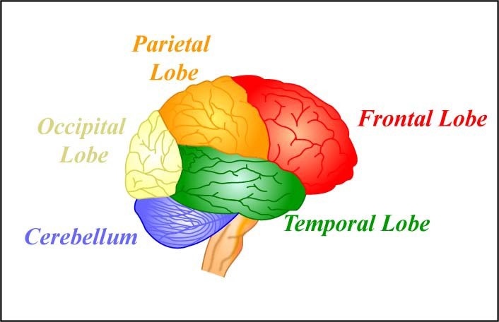
The four lobes of the brain and the cerebellum. [Image: MIT OpenCourseWare, https://goo.gl/RwUEVt, CC BY-NC-SA 2.0, https://goo.gl/Toc0ZF]
The temporal lobe is also involved in auditory processing, memory, and multisensory integration (e.g., the convergence of vision and audition). The parietal lobe houses the somatosensory (body sensations) cortex and structures involved in visual attention, as well as multisensory convergence zones. The frontal lobe houses the motor cortex and structures involved in motor planning, language, judgment, and decision-making. Not surprisingly then, the frontal lobe is proportionally larger in humans than in any other animal.
The subcortical structures are so named because they reside beneath the cortex. The basal ganglia are critical to voluntary movement and as such make contact with the cortex, the thalamus, and the brain stem. The amygdala and hippocampal formation are part of the limbic system, which also includes some cortical structures. The limbic system plays an important role in emotion and, in particular, in aversion and gratification.
