A Brain Divided
The two cerebral hemispheres are connected by a dense bundle of white matter tracts called the corpus callosum. Some functions are replicated in the two hemispheres. For example, both hemispheres are responsible for sensory and motor function, although the sensory and motor cortices have a contralateral (or opposite-side) representation; that is, the left cerebral hemisphere is responsible for movements and sensations on the right side of the body and the right cerebral hemisphere is responsible for movements and sensations on the left side of the body. Other functions are lateralized; that is, they reside primarily in one hemisphere or the other. For example, for right-handed and the majority of left-handed individuals, the left hemisphere is most responsible for language.
There are some people whose two hemispheres are not connected, either because the corpus callosum was surgically severed (callosotomy) or due to a genetic abnormality. These split- brain patients have helped us understand the functioning of the two hemispheres. First, because of the contralateral representation of sensory information, if an object is placed in only the left or only the right visual hemifield, then only the right or left hemisphere, respectively, of the split-brain patient will see it. In essence, it is as though the person has two brains in his or her head, each seeing half the world. Interestingly, because language is very often localized in the left hemisphere, if we show the right hemisphere a picture and ask the patient what she saw, she will say she didn’t see anything (because only the left hemisphere can speak and it didn’t see anything). However, we know that the right hemisphere sees the picture because if the patient is asked to press a button whenever she sees the image, the left hand (which is controlled by the right hemisphere) will respond despite the left hemisphere’s denial that anything was there. There are also some advantages to having disconnected hemispheres. Unlike those with a fully functional corpus callosum, a split-brain patient can simultaneously search for something in his right and left visual fields (Luck, Hillyard, Mangun, & Gazzaniga, 1989) and can do the equivalent of rubbing his stomach and patting his head at the same time (Franz, Eliason, Ivry, & Gazzaniga, 1996). In other words, they exhibit less competition between the hemispheres.
Gray Versus White Matter
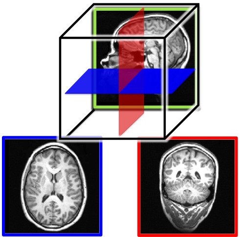
The cerebral hemispheres contain both grey and white matter, so called because they appear grayish and whitish in dissections or in an MRI (magnetic resonance imaging; see, “Studying the Human Brain”). The gray matter is composed of the neuronal cell bodies (see module, “Neurons”). The cell bodies (or soma) contain the genes of the cell and are responsible for metabolism (keeping the cell alive) and synthesizing proteins. In this way, the cell body is the workhorse of the cell. The white matter is composed of the axons of the neurons, and, in particular, axons that are covered with a sheath of myelin (fatty support cells that are whitish in color). Axons conduct the electrical signals from the cell and are, therefore, critical to cell communication. People use the expression “use your gray matter” when they want a person to think harder. The “gray matter” in this expression is probably a reference to the cerebral hemispheres more generally; the gray cortical sheet (the convoluted surface of the cortex) being the most visible. However, both the gray matter and white matter are critical to proper functioning of the mind. Losses of either result in deficits in language, memory, reasoning, and other mental functions. See Figure 3 for MRI slices showing both the inner white matter that connects the cell bodies in the gray cortical sheet.