
IV. STEPS TOWARDS CONSCIOUSNESS
32. THE HARD PROBLEM
In the 1640s Rene Descartes proposed that our awareness of the external world came from innate God-given abilities. (Science later substituted genetics for God – genetics whose accuracy was explained in terms of survival 200 years later by Charles Darwin.) Before that, in 1690, John Locke asserted that everything we know is information gained from our senses. Thus learning by observation became the academic basis of science. Subsequently, in 1710, George Berkeley explained that this learning involved associating sensory inputs with consistent experiences from various perspectives, other senses, and memory.
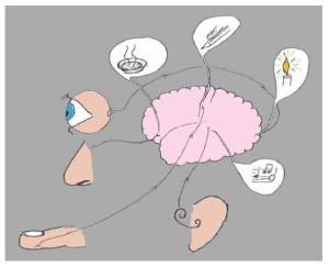
Despite a second Hundred Years War in Europe, the next century saw advances in anatomy and physiology. By 1838 Johannes Muller had found that all nerve impulses were the same whether they came from the eyes, ears, or skin. What determines their effect is where they arrive in the brain – his Doctrine of Specific Nerve Energies. [1]
For example, consider how the picture-in-our-head appears to be “out there”. From the brain’s perspective, the “out thereness” of the visual image is not substantially different from the “out-thereness” of what we experience when holding a pencil:
The pencil’s contact with our fingertips is sensed by pressure receptors in the finger tip. Their responses are conveyed by nerve impulses to specific locations in our somatosensory cortex. However, our immediate experience is not pressure on our fingertips. We feel the pencil’s hard surface at the fingertip’s location. In turn, that location is instantly calculated from limb angle receptors in the joints of our fingers, wrist, elbow, and shoulder. Even as we trace the nerve impulses from fingertip to the cortex, nothing about their arrival reveals their origin – except where they arrive.
Similarly, experiencing “a bright spot out there” when optic nerve impulses arrive at a specific location in the visual cortex is no more profound than experiencing those impulses from a finger as a “surface”.
There is something intrinsic about cortical locations that produces specific subjective effects.
These insights were consistent with later discoveries about the gradual development of various aspects of vision through infancy. “Development of the brain proceeds through a variety of stages in which the earliest events are programmed at the genetic level, while later ones are increasingly a function of neural activity, at first spontaneous in origin and finally resulting from environmental stimulation” Swindale (1996). [2]
For example, postnatal rats and other mammals raised in darkness for a critical period before being provided light never develop normal vision and behave as if blind. This has long been regarded as evidence that normal receptive fields require learning to form. Yet, dark-reared rats develop normal vision if given a neural growth factor hormone (NGF). [3] The researchers concluded that “ – NGF engages an innate program of visual development which is normally triggered by visual experience” (Pizzorusso, et al, 2000). Such innateness may be supported by some readers’ personal experiences of new-born infants attending to faces, which is consistent with recent fMRI data on the presence of facial, scene, and body selective locations in the infant cortex by 2.8 months. [4]
Developments in morphogenesis are beginning to answer these questions. [5] Cells become internally specialized as fetal developmental timing selectively activates genetic information. Simultaneously, they determine their location by exchanging chemical signals with adjoining cells. Such processes govern the visual system’s development, giving rise to the cortical maps with their receptive fields.
From retina to cortex, the visual system’s maps are intrinsic with their development from embryonic ectoderm (skin) tissues starting around 20 days after conception. [6] Our skin, itself is a map, a map of our body.
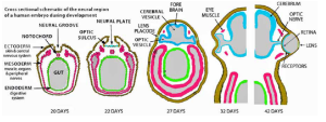 As the embryonic brain grows out from the neural tube, optic vesicles extend left and right towards the ectoderm. Contact instigates those ectoderm cells to form eye tissues, including the photoreceptors. That structure then folds inward, resulting in a ball with the photoreceptors in the back overlaid by brain tissue, which forms the bipolar and ganglion cells whose axons stretch back to the mid-brain. The retina is a part of the brain mapped by the same ectoderm mechanisms that shape our body and eyes. This mapping tendency then spreads centrally as the visual system develops.
It could be said that our visual system convert a body mapping ability to mapping the outside world.
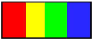
Another example of the innateness of visual processing arose when Hard & Sivak (1996) asked people to select colors that were pure red, yellow, green, and blue from a wide range of colors. The consistency of 80,000 results led them to conclude that color appearance had a genetic basis. These measurements led to a new standard system, the Natural Color System, for specifying color based on color perception. Consistent with this evidence, Bannert & Bartels (2025) recently report that different persons show similar patterns of cortical activity when seeing the same colors. [7]
A genetic basis for sensory qualities like color should not be surprising. The molecular structure of the long-, medium-, and short-wavelength receptor proteins are embedded in our genetic code. There is no question about the structure of our bodies being genetically determined. It is equally likely that certain aspects of behavior and cognitive processing are also determined genetically.
Since details of color, line, and shape depend on cortical location, could there be something particular about those neurons themselves? Growing evidence of the complexity within single neurons indicates sufficient complexity to encode processing as well as structure. [8]
Nevertheless, observations like these still leave the Hard Problem unanswered.
- Mueller’s Doctrine is routinely demonstrated during surgery to mitigate the effect of epileptic seizures. Due to individual differences in human brain anatomy, the surgery requires direct testing to determine the exact location of specific functions. This is done by probing the cortical surface with mild electric pulses, asking the patient what they sense, and looking for reflex movements. (The patient must be awake, but this is not a problem since external brain stimulation does not produce pain. Only specific neural pathways do that.) Under local anesthetic, part of the skull is lifted so mild electric pulses can prod the cortex to map out those particular functions. Notably, identical pulses produce different effects depending on where they are delivered. ↵
- Swindale NV (1996) The development of topography in the visual cortex. Computation in Neural Systems, 7, 161-247, p. 233. ↵
- Pizzorusso T, Fagiolini M, Gianfranceschi L, Porciatti V & Maffei L (2000) Role of neurotrophins in the development and plasticity of the visual system: experiments on dark rearing. International Journal of Psychophysiology, 35, 189-196, p.191 {doi.org/10.1016/S0167-8760(99)00053-7}. ↵
- Kosakowski HL, et al (2022) Selective responses to faces, scenes and bodies in ventral visual pathway of infants. Current Biology, 32, 265-274 {doi.org/10.1016/jcub.2021.10064}. ↵
- Markovic A, Briscoe J & Page KM (2024) Dynamics of positional information in the vertebrate neural tube. BioRxiv, 6 Sept., 1-9 {https://doi.org/10.1101/2024.07.01.601514} ↵
- The figure is based on S. Polyak’s The Eye, University of Chicago Press, 1957. See Saade M & Marti E (2025) Early spinal-cord development: from neural tube formation to neurogenesis. Nature Reviews Neuroscience, 26, 195-213 {doi:org/1038/s41583-025-00906-5}.That visual mapping is derived from skin mapping is suggested by our ability to visualize proprioceptive location of our hands and feet with our eyes closed: Close tour eyes and touch your nose with a finger tip. Such visual mapping outside our skin would have been a major asset in making and using tools – our major defensive against against better armed predators. ↵
- Hard A, Sivik L & Tonnquist G (1996) NCS, Natural Color System - from concept to research and applications. Part 1. Color Research and Application, 21, 180-205. Bannert MM & Bartels A (2025) Large-scale color biases in the retinotopic functional architecture are shared across human brains. Journal of Neuroscience, 38, 3657-3668. ↵
- Nehme R, Pietilainen O & Barret LE (2024) Genomic, molecular and cellular divergence of the human brain. Trends in Neurosciences, 47, 491- 505 {doi.org/10.1016/j.tins.2024.05.009}. Bonev B (2024) Opportunities and challenges of single-cell and spatially resolved genomics methods for neuroscience discovery. Nature Neuroscience, 27, 2292-2309 {doi.org/10.1038/s41593-024-01806-0}. For a delightful introduction to the complexity of single cells, see: Bray D (2009) Wetware: A computer in every cell. Yale University Press. {https://www.jstor.org/stable/j.ctt1np9f2.2}. ↵