
APPENDIX F – Directing Your Gaze
For the visual system to know where we look and obtain distance information from the accommodation and the vergence processing, it must be able to control eye muscles accurately. (Appendix G – How Muscles Work explains how the brain can make muscles contract in response to nerve impulses.) Most muscles in our body are controlled by feedback from stretch receptor cells in a muscle and from pressure sensors in the limb joints. This is too slow and not precise enough to control eye muscles, which therefore lack such receptors. Eye muscles are controlled directly by the brain based on the visual image. That image processing is enhanced by some front-end neural networks that streamline the control signals from the visual cortex.
GAZE DIRECTION
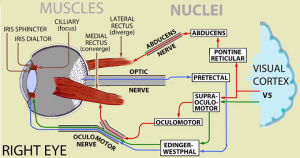
To control where we look, we use two neuromuscular mechanisms in an alternate manner:
The right eye’s medial rectus muscle is linked to the left eye’s lateral rectus. Stimulation of this neural net turns both eyes towards the left.
Another net links the right eye’s lateral rectus to the left eye’s medial rectus muscle. Stimulation of this net turns both eyes towards the right.
The balance of simultaneous activation of these two networks precisely controls where we look.
FOCUS
The focal length of the lens in our eye determines the distance from which a shape is sharply focused on the retina, 25 mm behind the lens. (See Ch 24.) In its relaxed state, our eyes are focused at a distances of 3 meters (10 feet) and beyond. To look closer, the lens’ curvature must be increased to bend the incoming rays more and focus them on the retina. The lens in our eye is flexible and will bulge if squeezed around its edge. This is done by contracting the ciliary muscles. The impulses from the visual cortex, via the supraoculomotor and Edinger-Westphal nuclei, adjust their contraction to maximize the high spatial frequencies in the image of the shape.
When a shape is focused, feedback from the rate of these impulses informs the visual system of the shape’s distance.
VERGENCE
Linked stimulation of the medial rectus muscles in both right and left eyes increases convergence of gaze, while linked stimulation of the lateral rectus muscles in both eyes increases their divergence. A maximum strength of low spatial frequencies in their combined visual image indicates the retinal images in each eye fully overlap.
The relative strength of the convergent and divergent signals tells us the shape’s distance.
PUPIL REFLEX
While the above are controlled by analysis of the visual image, the basic control of pupil size does not come from the visual cortex. Secondary pathways from the retina directly to the pretectal and Edinger-Westphal nuclei operate the iris sphincter muscle to vary the degree of contraction in response to the general light intensity of the retinal image.
Simultaneously adjusting the intensity of the retinal image as we look in different directions enables maximizing lightness contrast to make the high and low spatial frequencies more evident. See for example, the crispening effect in Ch 17.
The brain’s level of arousal influences the responsiveness of all these mechanisms. Because the pupil reflex is somewhat independent of the visual cortex, its response to strong emotions such as fear and love can be evident. [1]
- Joshu S & Gold JI (2020) Pupil size as a window on neural substrates of cognition. Trends in Cognitive Sciences, 24, 466-480; https://doi.org/10.1016/j.tics.2020.03.005. ↵