
II. FROM OPTICAL TO VISUAL EFFECTS
12. FROM PHOTONS TO NERVE IMPULSES
Neurons and all the cells in our body live in a salty intercellular bath that mimics the early ocean in which life first evolved. We continue to bear that ancient ocean within us to sustain the original mechanisms that support life.
All animal cells have a skin (membrane) made of a double layer of fatty molecules. The cell membrane must be semipermeable to ions and small molecules. Oxygen, water, and nutrients must be able to enter, while CO2 and waste need to leave. This membrane also holds complex protein molecules that selectively admit nutrients, remove waste products, release substances (like neurotransmitters and digestive enzymes), extend structures (like hair and links to other cells), or react to specific substances, to pressure, and to photons.
Inside these cells, proteins carry on the cells’ functions. Dissolved in the intercellular fluid, these proteins ionize and acquire a negative charge by holding on to otherwise shared electrons. That gives the cells an inside charge of minus 70 millivolts. Therein lies a problem:
Surrounded by a salty body fluid with a relatively high concentration of dissolved Na+ (sodium) and Cl– (chlorine) ions, that internal -70mV is highly attractive to the Na+ ions. These sodium ions constantly leak through the cell’s membrane. Chemical calculation (the Nernst Equation) indicates they would continue to enter until the cell reaches +40 millivolts. Thereupon, the cell would soon die as its internal functions cease.
(Shortly we shall see how this vulnerability has proven to be useful.)
To maintain their -70 mV, cells operate pumps that continually remove the in-leaking Na+ ions. Indeed, every cell in our body has thousands of such pumps in its cell membrane. This is a lot of bother, but evolution has turned it into an advantage in neurons by using the sodium ions’ electrical potential for communication with other neurons.
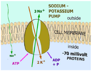 Sodium-potassium pumps push 3 Na+ ions out in exchange for 2 potassium, K+, ions brought in:
This requires energy. It is obtained by splitting a molecule of adenosine triphosphate (ATP) into adenosine diphosphate (ADP) and a phosphate molecule.
Mitochondria (the powerhouse structures in cells) burn glucose to obtain the energy for recombining ADP and phosphate into ATP. Without a continuous supply of glucose for the mitochondria, cells soon die when the pumps fail to maintain the interior at -70mV.
Photoreceptors
Photons do not trigger the photoreceptor cells to emit nerve impulses. They do not even provoke them to release neurotransmitters. (See Appendix C – Neurotransmitters.) Rather, the visual system uses reverse logic:
Photons cause the photoreceptors to stop releasing glutamate. This neurotransmitter then stops bipolar neurons from exciting ganglion neurons whose axons form the optic nerve.
Why this indirect approach? You may have heard the expression, “The quick and the dead”. It can be easier to stop things than to get things going. Having the receptors continuously active in darkness enables photons to produce an effect by blocking the photoreceptor release of glutamate.
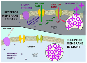
In darkness, photopigments in the receptor cell membrane continually activate a recurrent chain of events that releases messenger molecules. These open sodium gates. The inflowing positive sodium ions raise the receptor’s potential from -70 to -40 mV. This voltage opens calcium gates. Inflowing calcium ions release the glutamate neuro-transmitter:
In light, absorption of a photon changes the photopigments so they no longer activate the release of messenger molecules. The sodium gates close. Sodium-potassium pumps restore -70 mV within a millisecond. The calcium gates close. Lack of calcium stops the glutamate release:
Neural Connection
The photoreceptor’s glutamate is received by two types of bipolar neurons having either inhibitory or excitatory synaptic receptors. This makes them selectively sensitive to light or dark spots in the retinal image. [1]
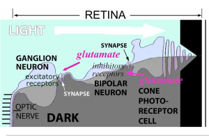 Here is how a bipolar cell with inhibitory glutamate receptors responds in light and in dark:
In light, the photons stop the photoreceptor from releasing glutamate. Consequently, the bipolar neuron is no longer inhibited. It releases gluta- mate to a ganglion neuron which has excitatory glutamate receptors. The ganglion neuron responds by releasing impulses into the optic nerve.
In dark, the photoreceptor releases glutamate. This inhibits the bipolar neuron from releasing glutamate to the ganglion neuron. Consequently, the ganglion cell rarely releases nerve impulses.
Conversely, a bipolar neuron with excitatory glutamate receptors only responds when the photo-receptor releases glutamate in darkness. Therefore, its ganglion neuron is only excited in darkness.
Responding to dark parts of a retinal image is as important as responding to the bright areas – as demonstrated by your reading these words.
Nerve Impulses
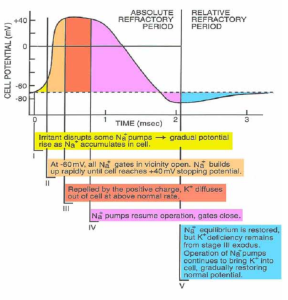
Nerve impulses are brief pauses in a neuron’s “activity of daily living”. Neurons differ from other cells in having a cell membrane with molecular sodium ion (Na+ ) gates that they normally hold closed. Irritating a neuron distracts it, so some of these gates open. The inflow of these positive ions raises the neuron’s voltage above -70 mV. If irritated sufficiently that enough gates open and the voltage increases to above -60 mV, a drastic change occurs:
All Na+ gates open, and Na+ ions rush in until their concentration balances the attraction of negative protein ions. This balance is reached at +40 mV. The neuron would then die unless it quickly recovers and closes the Na+ gates. As the sodium pumps resume operation, the voltage inside the neuron drops. The nerve impulse is that millisecond of positive voltage.
Until the pumps have removed the excess Na ions, the neuron is in an absolute refractory state – no amount of irritation will open its sodium gates. In this state, the voltage is well below normal -70 mV because many potassium ions (K+) left when the internal voltage was +40 mV.
The K+ balance is gradually restored as the sodium pumps deal with continual Na+ leak by exchanging 3 Na+ ions for 2 K+.
During this restoration, the neuron is in a relative refractory state, and strong irritation will open some Na+ gates. As the voltage returns to -70 mV, less irritation suffices. This enables neurons to encode the strength of irritation by how soon they can produce another impulse.
The 2-millisecond absolute refractory state limits the repetition rate to about 50 Hz.
Neurons are not as stable as the above may imply. They typically fire at random once every ½ to 2 seconds. (Local fluctuation in ion concentrations and availability of ATP energy molecules to power the pumps are likely culprits.) This reduces the dynamic signal range to as low as 1 to 25 times the random level. Nevertheless, there is a significant advantage:
A neuron that releases no impulses for 2 seconds or more signals that it is being actively inhibited. Our nervous system has been regarded as similar to digital computers because its nerves either fire or not, and their impulses are always the same intensity and duration. However, it is actually a trinary system. Its three states (firing, random, and inhibited) enable considerably more complex and efficient information coding.
Impulse Conduction
That neurons can produce impulses and recover from a temporary breakdown is of little value if that information doesn’t go somewhere. During the height of the impulse, the concentrated Na ions quickly spread to an adjacent region of the neuron. As the voltage there rises above -60 mV, a nerve impulse appears. And so forth:
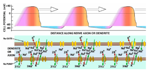
The absolute refractory period prevents backfire, ensuring the impulses spread in one direction.
Because more ions spread faster, impulses spread faster in large axons and dendrites. Their spread is diminished by leakage through the cell membrane. The larger surface-to-volume ratio in small fibers slows their impulses. Small neural axons and dendrites vary in diameter from 0.2 to 2 μm, resulting in impulse speeds from 0.5 to 2 m/sec. However, stopping the leakage enables the ion concentration to spread further. This is accomplished by glial cells’ myelin wrapped around nerve fibers. This coating increases the speed 10X. Larger nerve fibers up to 20 μm diameter are usually coated. Impulses in these can travel over 100 m/sec. Fortunately, or else we would be pretty slow thinkers and movers.
Synapses
Neurons send impulses from their cell body along a tubular extension of their membrane, an axon. They receive stimulation from other neurons’ axons either directly or to an extended membrane “arm”, a dendrite. Neurons have only one axon, which may branch. Neurons may have many dendrites with many branches. Nerve impulses appear to pass from neuron to neuron even though they are not connected.
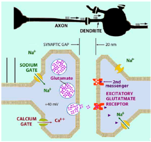 There is always a gap, synapse, between axons and dendrites. These interruptions between neurons enable neural systems to do more than just relay information.
Axon-dendrite synapses are similar to those between photoreceptors and bipolar cells. When a +40 mV impulse arrives at the end of an axon, it opens calcium gates. The inflow of Ca++ ions drives neurotransmitter packages (vesicles) to the end membrane. There they release their contents into the synaptic gap.
It takes 0.5 milliseconds for these molecules to diffuse across the 20 nm gap. Whereupon they may enter a receptor in the dendrite membrane. The receptor reacts by directly opening a sodium gate or releasing 2nd messengers, to open such gates. As Na ions enter, the dendrite voltage rises producing a nerve impulse.
(Glutamate is used extensively in the visual system.)
- It is difficult to illustrate that: 1) The cones continuously respond in darkness by releasing the neurotransmitter glutamate in their synaptic connections to the bipolar cells. 2) The cones stop responding in light, which also explains rapid light adaptation. 3) The bipolar cells also release glutamate into their synapses with the ganglion cells. 4) Glutamate can have either an excitatory or inhibitory effect depending on the receiving receptor in the synapse. 5) Both cone and rod photoreceptors also interconnect by non-synaptic gap junctions. (Not shown are horizontal and amacrine cells that synaptically interconnect adjacent bipolar and ganglion cells. These cells are involved in dark adaptation and acuity.) ↵