
II. FROM OPTICAL TO VISUAL EFFECTS
24. AN ENORMOUS ACUITY PROBLEM
Lateral inhibition sharpens acuity by overcoming the effects of scattered light. However, there is a more serious barrier to sharp visual images:
Some 100 million receptors sense the directions from which photons arrive, but the optic nerve has only a million ganglion cell axons.
How can we see details as small as a single receptor?
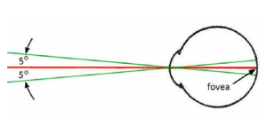 The visual system’s solution only gives us good acuity over a 5 degree angle to either side of straight ahead. That comes from some 6 million cone receptors in the clustered in center of the retina – the fovea. About 5 cones connect to each optic nerve axon – with overlap. This results in small, wavelength-sensitive receptive fields for each axon.
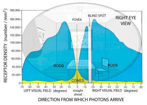
Outside the fovea, the density of cones decreases rapidly – replaced by some 100 million rod receptors. Up to a hundred or more of these connect to a single optic nerve neuron – greatly increasing its sensitivity as scotopic vision, but with a corresponding loss of acuity.
With a little effort you can experience this limited vision yourself:
* Pick some spot in front of you.
* Gaze steadily at that spot. (You may find this easier by closing one eye.)
* Attend to something left or right of that spot without moving your eyes.
* Notice how what is further out is not sharply focused and that colors also fade.
This figure shows the distribution of these two receptor types across the retina:
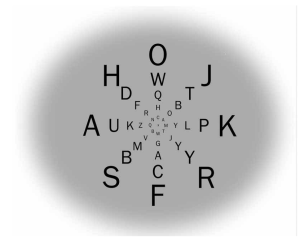
This figure shows how acuity decreases for targets further out from your view straight ahead:
Relative to the center, these letters increase in size relative to the decrease in acuity from the center to the periphery of vision.
Viewed from a certain distance, all letters are equally legible.
We are usually unaware of this limited acuity because we keep shifting our gaze to see other details around us.
The visual system uses memory to fill in details as we gaze elsewhere.
80% of what we see is memory!
This is possible because short-term visual memory for details of shape and color is excellent over many seconds.
Still, where to fill-in those details requires precise knowledge of eye position and control of eye movement. The precision required to fill-in and assemble an image forced the visual system to evolve a unique solution to direct and record eye position rapidly:
Eye muscles are unique. There is no feedback from stretch receptors. That would be too slow. Instead, the visual system monitors the movement of the visual image and directly adjusts the muscles. The feedback control is image guided.
(To obtain accuracy and overcome inertial error, analog motion control systems introduce a small positional jitter that is constantly corrected by feedback. The average of these jitters is more precise than direct positioning.) Visual systems evolved this technique long ago. In the eye this jitter extends over a few receptors, too small to be noticed – microsaccades. (This also prevents the picture-in-your-head from fading during fixated staring by constantly shifting the retinal image back and forth. This prevents specific receptors from exhausting their resources.)
Appendix F- Directing Your Gaze describes the immediate neural mechanisms that control the direction and distance at which the eyes are focused.
Appendix G – How Muscles Work explains how nerve impulses can make muscles contract – the basis of every movement you make.
Here is a powerful demonstration of how the visual system “fills in” an image when our gaze is elsewhere:
In the figure of the retina in Ch 24, at the viewing direction 15 degrees to the right in the right eye, there are no receptors. This is where the optic nerve leaves the eyeball’s interior to reach the rest of the brain. It is labeled “BLIND SPOT” because the lack of receptors means the image there can not be seen.
There is a corresponding region towards the left in the left eye. What we usually see is a composite of the views from both eyes. We are not aware of either blind spot because the image in the blind spot of one eye is covered by that view in the other eye.
To see your blind spot, copy this diagram on a white card, placing a similar size X and a little face about 10 cm (4 inches) apart.
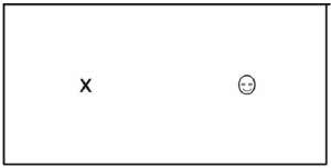
Cover your left eye. Stare directly at the X from about 30 cm (12 inches).
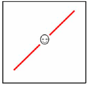
Your visual image is not empty in the direction of the blind spot. To see that the visual system actually fills-in the blind spot rather than collapsing or leaving it blank, repeat the above after drawing a diagonal red line across the face, but keep the line out of the face itself. [1]
- The visual system “fills-in” the blind spot to avoid spatial distortion in the overall picture. The completed line even though “interrupted” by the blind spot indicates the visual system does more than simply extend the background. It uses information from a substantial portion of the picture to strengthen a segment of uncertain detail. The two figures on pages 90 and 91 were other examples of this processing. To learn more about the blind spot, see Spillmann L, Otte T, Hamberger K & Magnussen S (2006) Perceptual filling-in from the edge of the blind spot. Vision Research, 46, 4252-4257. To learn more about the role of microsaccades in short-term memory, see Quan G, et al (2024) Microsaccades reflect attentional shifts: a mini review of 20 years of microsaccadic research. Frontiers in Psychology, 19 February, DOI: 10.fpsyg.2024.1364939. ↵