
III. ASSEMBLING THE VISUAL IMAGE
26. IMAGE NEURONS
A visual image could be formed by connecting specific line- and angle-sensitive neurons to a neuron that would then respond to just that image. Even so, you might also be skeptical. There are so many images and potential images for us to see that even 16 billion neurons in the cortex would be nowhere near enough. [1] Yet there is evidence that the visual system has image-sensitive neurons, to a limited extent.

A visual illusion contributed towards this discovery in 1980. Originally noted in a doctored photograph of Britain’s Prime Minister, Margaret Thatcher, it has been named accordingly: [2]
The left pair of faces look normal, whether or not inverted. We see nothing wrong with the inverted face on the right, but non-inverted below, it looks horrid. This suggests that we have an innate ability to recognize faces. Such genetic “tuning” enables rapid correction and identification of faces when they are viewed from various perspectives.
This possibility was supported by studies that had found human infants preferred looking at faces within days after birth.
In 1982, David Perrett, Edmund Rolls, and Woody Caan made micro-electrode recordings from single neurons in the temporal lobe of monkeys, and found neurons that responded selectively to faces. [3]
52 1997, non-invasive (magnetic resonance imaging – MRI, see Ch 30) enabled recording visual responses at specific human brain locations. Nancy Kanwisher, Josh McDermott and Marvin Chun found face- and shape-sensitive single neurons in the lateral and inferior regions of the occipital and temporal lobes. [4] The green outlined location in the left hemisphere is where the stronger responses to faces (F) than objects (O) occurred, as shown in the graph on the right:
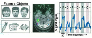 In 2023, Quiroga, et al obtained face selective responses from single cells in this area of human brains. These findings explain why damage in these areas can result in prosopagnosia – an inability to recognize faces.
What and Where
Various studies with humans and monkeys find other locations in the ventral-lateral areas of the occipital and temporal lobes that respond to shapes, such as body parts, letters, and certain aspects of natural scenes. These descending connections from the primary visual cortex are called the WHAT channels. Long neural tracts to and from the frontal lobe support their role in recognizing these shapes.
By contrast, upward connections from the primary visual cortex into the dorsal occipital, medial temporal, and lower parietal lobes areas are called the WHERE channels. Responses in these areas are related to location in the retinal image and also to movement. [5]
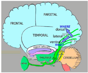
Area V1 (where the optic nerve tract
connects to the cortex) and areas V2 –
V4 are located on the rear cortical surface and into the medial surface between the two cerebral hemispheres. Connections between these areas, from them to the dorsal, lateral, and ventral regions of the visual cortex, and to the temporal, parietal, and frontal lobes are all subcortical – not along the surface.
The cerebral cortex is actually a 2.2 meter2 sheet of neural tissue, only 3 mm thick. It is folded and crumpled to fit inside our skull. The space inside is filled with long, myelin wrapped axons and dendrites connecting various locations in that sheet, and connecting it to various neural structures within, such as the thalamus.
- Stent G (1975) Limits to the scientific understanding of man. Science, 187, #4161, 1052-1057 ↵
- Thompson, P. (1980) "Margaret Thatcher: a new illusion". Perception. 9 (4): 483–484 {doi:10.1068/p090483. PMID 6999452. S2CID 32492890}. ↵
- Perrett DI, Rolls ET & Caan W (1982) Visual neurones responsive to faces in the monkey temporal cortex. Experimental Brain Research, 47, 329-342. ↵
- Kanwisher N, McDermott J & Chun, MM (1997) The fusiform face area: A module in human extrastriate cortex specialized for face perception. Journal of Neuroscience, 17, 4302-4311, fig. 3a. Quiroga RQ, et al (2023) Single neuron responses underlying face recognition in the human midfusiform face-selective cortex. Nature Communications, 14, 5661,{doi.org/10.1038/s41467-213-41323-5}. ↵
- Ungerleider LG & Haxby JV (1994) “What’ and “Where” in the human brain. Current Opinion in Neurobiology,4, 157-165. Ayzenberg V, Simmons C & Behrmann M (2023) Temporal asymmetries and interactions between dorsal and ventral visual pathways during object recognition. Cerebral Cortex Communications, 4, 1-12. ↵