
III. ASSEMBLING THE VISUAL IMAGE
28. EXTRACTING DISTANCE
After determining the directions from which certain photons have arrived, the most profound ability of the visual system is its ability to sense the distance of their source. This quality of sensing things far beyond our body makes the “picture-in-our head” so valuable for survival. Several processes extract distance information from the flat retinal images:
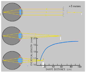
Accommodation
The visual system must change the eye’s focal length to see shapes sharply at various distances. Contracting the muscles around the circumference squeezes the flexible lens, increasing its curvature:
How does the system know when an unknown shape is in focus?
As an image sharpens, its high spatial frequencies increase in strength. Fourier Analysis measures the high-frequency component of the retinal image. Feedback adjusts the lens to maximize that component. The stronger the signal squeezing the lens, the closer the shape.
This reflex signal alone is accurate only up to a meter. Yet this short-range accuracy is useful for vital work such as picking ticks and knapping stones.
(This is another example of feedback control – like the one for brightness on p. 65.)
Binocular Vergence
A shape looks sharpest when it is focused on the fovea. As a shape comes closer, each eye must rotate inwards – converge to keep its image on the fovea. How much the eyes converge reflects the shape’s distance.
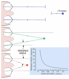
This graph shows how convergence changes with distance. The vergence angle refers to how much the eyes rotate inwards from looking straight ahead.
Similarly to how it controls lens focus, the visual system uses Fourier Analysis to measure sharpness of the retinal image.
When the left and right visual images overlap, low spatial frequencies maximize. Feedback adjusts the eye muscles to maximize the image’s low frequencies.
The vergence signal sent to the muscles changes little when a shape is further than 2 meters. Yet that distance range is valuable for grasping or ducking branches when racing through trees. Go to APPENDIX F – Directing Your Gaze to see how the visual system operates the focus and vergence muscles.
Binocular Disparity
Another distance sensing process uses the difference, disparity, in the two eyes’ retinal images when focused on the same shape. See this disparity yourself by holding up a finger 10 centimeters from your nose. Look at the finger with one eye and then the other. Now, straighten your arm and repeat. Notice how much less the finger moved relative to the background at arm’s length. The images in the two eyes differ less for distant shapes. [1]
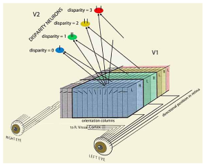
The rows of edge and line orientation visual-field columns in V1 of the visual cortex have alternate inputs from the left and right eyes. In turn, successive pairs of rows have inputs from adjacent positions in the retina. These neurons connect to disparity neurons in area V2.
The disparity between left and right images decreases with distance. Based on which disparity neurons are most active for a shape, the system places it at that distance in the visual image.
Disparity distance judgments are accurate about as far as a person can leap. However, adding accommodation and vergence information improves the judgment of distances out to 40 meters [2]
Disparity processing is also the basis of an additional effect produced when the visual system combines the two images into a single view of the world beyond our eyes – stereopsis. Its distinctness from seeing distance is analogous to seeing a nail as sticking up out of a board in addition to seeing its head as closer to you than the board. (From 5% to 20% of otherwise normally sighted persons lack stereoscopic vision. [3] )
Figure / Ground Perception
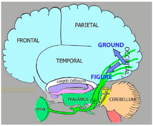
As the distances of various shapes in the retinal image are determined, attention focuses on one shape as the figure and relegates the rest to its background.
The figure information is then further processed in the ventral (lower) region of the occipital lobe and adjoining temporal lobe to identify the figure.
The background infor-mation is processed in the occipital lobe’s dorsal (upper) region and adjoining parietal lobe to determine the figure’s location.
These projections correspond to the WHAT and WHERE pathways described earlier in Ch 25.
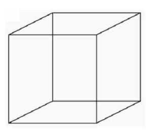 The tendency to regard a closer shape as the figure and the remainder as background is evident in this drawing:
Attend to the two inner vertical lines. Select the one to the right, and you see the cube from above while the left line recedes to the background. Select the left line, and you see the cube from below. (This may take a moment.)
We can’t see both views simultaneously because focusing on one line directs each line’s information to different areas of the visual cortex. The speed at which you can switch your attention between the lines and change your image of the cube indicates complete maps of the retinal image exist in both dorsal and ventral regions.
Separate processing of figure and ground organizes the visual image to avoid confusion.
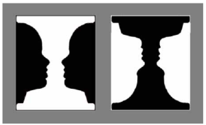 These images demonstrate that our thoughts influence what becomes figure or ground:
Faces predominate on the left because we are attentive to faces and tend to see dark surfaces as closer.
The vase dominates on the right because it is dark, and the faces are inverted.
Yet, deciding on a white vase or white faces, can reverse figure and ground. [4]
- If you encounter a bear in the wild, you may see it raise up on hind legs and sway back and forth. Bears have poor acuity. Yet they `know` about the relation between distance and retinal disparity. By swaying back and forth, the bear increases the difference between the retinal image when leaning left and the image when leaning right. The greater your image moves against the background, the closer it judges your position. You can reduce the disparity by swaying in the same direction as the bear. Thereby, you increase your apparent distance and, hopefully, your threatening appearance. ↵
- Palmisano S, et al (2010) Stereoscopic perception of real objects at large distances. Journal of Vision, 10, 1-16. ↵
- Levi DM (2022) Learning to see in depth. Vision Research, 200, {https://doi.org/10.1016/j.visres.2022.108082}. ↵
- Evidence that background information is also used to identify figures was found in a series of experiments on teaching reading skills in kindergarten. Children learning to read have reported that the letters “all look alike”: Nelson TM, et al (1999) added information in the background of individual letters of the alphabet to see if making them more distinct would help (Improving perception of letters and visual structure of language. Perceptual and Motor Skills, 88, 515-530.) Compared to children with low reading skills trained with plain and different textured letter backgrounds, those trained with patterned backgrounds significantly improved their skills. For further insight into figure-ground processing, see van der Hyedt R (2015) Figure-ground organization and the emergence of proto-objects in the visual cortex. Frontiers in Psychology,6, article 1695 {doi/10.3389/fpsyg.2015.01695i}. ↵