
II. FROM OPTICAL TO VISUAL EFFECTS
22. WHENCE PHOTONS CAME
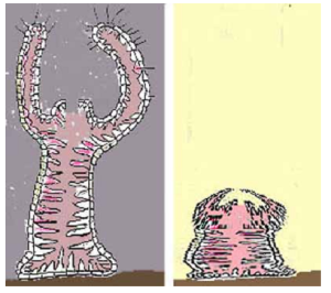 Just having some photoreceptors enables an animal to respond selectively to surrounding light conditions.
For example, in darkness, simple sessile animals such as tunicates open and wave their arms to capture food. In light, it is safer to hunker down and be inconspicuous:
Having two sets of receptors at different locations opens a world of possibilities for mobile animals. Comparing the receptors’ responses indicates the direction of the most or least light – a primitive sense of where stuff is located outside the organism. This information can then guide the animal’s behavior when combined with some instinctive or learned reactions.
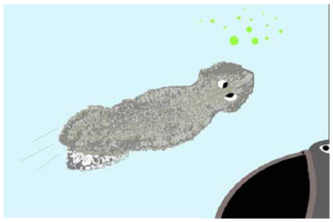
Neural connections from two clusters of photo-receptors to contractile tissue along its body enable this planaria worm to avoid darkness to its right and approach greenish light from algae at left.
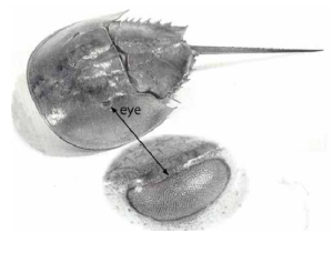
Arthropods, like horseshoe crabs, have external eyes that do not produce an optical image. Yet they can still tell whence photons came.
A photoreceptor on a curved surface receives the most light from a specific point in the direction faced by that receptor. Since other rays from that point will not effectively enter adjacent receptors, they are lost. That makes each receptor directionally selective.
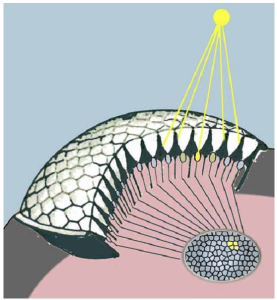
Such eyes face a major tradeoff between acuity and sensitivity. Smaller receptors respond to a narrower range of directions but gather less light.
Sorting the responses from an array of such receptors to different locations in a neural network amounts to a map of the directions from which they arrived.
Whether a visual “image” exists in that network is a matter for discussion elsewhere.
A lens focuses all available rays from any specific direction to a point in the image. Therefore, the receptors in that image can be as small as possible to maximize acuity. Creating an optical image of the source inside the organism protects receptor cells from the environment and stray light,
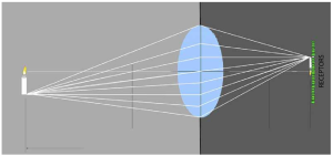 With such advantages, no wonder most vertebrates, some mollusks, and even some earthworms have evolved light-sensing organs using a lens to sort photons in terms of whence they came into an optical image inside an eye. An array of photoreceptor cells in that image maps the directional information into distinct nerve fibers that transmit the information for processing.
This schematic of the human eye shows how a receptor’s size (2.5 μm) determines its directional acuity or visual-field based on the eye’s focal length (25 mm) and the receptor’s distance from the lens (25 mm): That 22 second angle receptor field-of-view equals the width of a human hair at arm’s length!
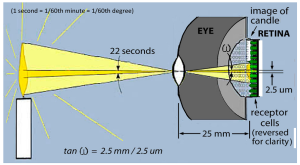 N.B. This figure uses a simplified retina to emphasize the basic optics. In vertebrate eyes, the photo-receptors face away from the lens. They are overlaid by bipolar, ganglion, and other neural cells, plus the blood vessels that sustain them. Light must travel through all these layers and the receptor cell body before reaching photo-sensitive pigments. Fortunately, these structures are nearly transparent. See more accurate figures in Ch 20 and 21.
Only vertebrate eyes use such a backwards design. Why? That’s also another story. [1]
- As shown in the figure on page 76, the receptors are furthest back in the retina. The vertebrate eye is organized in this manner because its neural structure is a direct extension of the brain. This does result in some loss of acuity, but processing is also necessary at this stage to connect the 100 million receptors to one million fibers of the optic nerve. While our photoreceptor cells are derived from epidermal tissue like the receptors for touch, taste, etc., the rest of the vertebrate retina is an outgrowth of the brain. This is evident at the earliest stage of embryo brain development. See page 156, and Fritzsch B & Martin PR (2022) Vision and retina evolution: How to develop a retina. IBRO Neuroscience Reports, 12, 240-248. We saw above the complex retinal processing involved in creating 6 types of color pathways from three types of receptors. Therefore, it is not surprising that brain abnormalities can impair such processing: Vidal KSM, Decleva D, Barboni MTS, et al, (2022) The association between acquired color deficiencies and PET imaging of neurodegeneration in mild cognitive impairment and Alzheimer Disease. Investigative Ophthalmology, and Visual Science, 63, 20. The ability to directly observe the brain through the pupil with a microscope provides a non-invasive means for neurological examination with potential clinical applications. For example: Shu, et al (2023) Retina oculomics in neurodegenerative disease. Annals of Biomedical Engineering, 51, 2708-2721 {https://doi.org/10.1007/s10439-023-03365-0} ↵