
II. FROM OPTICAL TO VISUAL EFFECTS
20. ANOTHER PROBLEM WITH PHOTONS:
They’re Not Colored!
The surfaces we see have no color, nor do the photons they reflect. For many people, this is their first clue that the picture-in-their-head differs from the physical world. For this reason, we shall look closer at how the visual system adds color to what we see. This examination is possible because, in many respects, more is known about how the visual system processes the effects of different wavelengths to produce color than is known about any other aspect of consciousness.
Perhaps the most convincing evidence that color is not an inherent characteristic of photons is that photons of a single wavelength produce different hues when pulsed for differ in duration and different rates. [1] These effects indicate that color perception depends on neural impulse patterns as well as which cones absorb the photons.
Consider, “How could the wavelength information available from photons be made evident in a conscious representation?” The image in our head is already filled with the finest possible directional detail available from the retinal image based on various densities of arriving photons. Adding some kind of symbol to indicate wavelength point by point would interfere with the image detail. Alternatively, adding some temporal effect like ”twinkling” would take time, thereby reducing the speed of perception and the ability to notice change.
Evolution hit the same solution physicists use when they can’t explain an idea: Invent a new “dimension”, such as strings:
Like the effect of brightness, color is an additional subjective dimension in the visual image to represent the wavelength of photons.
The pigment molecules that selectively absorb a specific range of wavelengths are genetically defined. Some 60,000 measurements found that people are very similar and consistent in selecting color samples that matched their idea of a perfect red, yellow, green, and blue from hundreds of samples that were barely distinguishable in hue. This indicates that these subjective dimensions are in our genetic code. [2]
Because photons are not colored, our brain has to do it. The following explains how it does that.
Differences in response strength from the three types of cones are processed to produce some 1/4 million colors. This begins in the retina. In 1966, DeValois, Abramov & Jacobs found that six types of optic nerve fibers carry color information to the cortex in opponent-color excitatory-inhibitory pairs: [3]
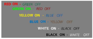
These names refer to how the color is affected by activity in that neural pathway.
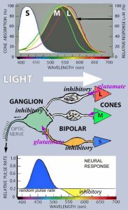
The following figures show simplified neural models of how the retina produces these color pathways.
Their intent is to explain these basic aspects:
1. Why the wavelength absorption peaks of the Long, Middle, and Short wavelength cone pigments do not match the wavelengths that produce the strongest red, green, and blue colors. This alone suggests that there is more than a simple transmission of wavelength information.
2. How yellow enters the picture as a 4th primary color – even though there are only three types of cones.
3. How a white pathway arises from a combination of all three cone responses, and black arises from reverse inhibition when there is no light.
As you work through the following schematics, remember (Ch 12):
a. Cones emit glutamate in darkness and cease doing so in light.
b. Bipolar neurons release glutamate unless they are inhibited.
c. There are two types of synaptic receptors for glutamate – one inhibits its
neuron, the other excites it.
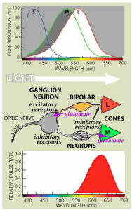
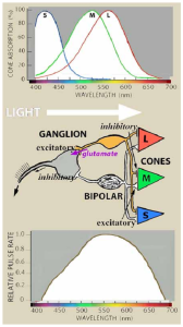
Red On – (Green Off Pathway)
The ganglion cell for this pathway is connected to two bipolar neurons. One bipolar excites the ganglion cell and has an inhibitory synapse with a Long wavelength cone. This cone absorbs light from 400 to 700 nm with a maximum at 560 nm. The other bipolar inhibits the ganglion cell and has an inhibitory synapse with a Mid wavelength cone that maximally absorbs 530 nanometers.
Because the L cone’s sensitivity is overlapped by the M cone’s beginning at 550 nm, the L cone’s bipolar excitation of the ganglion neuron gradually overcomes the M cone’s inhibition. Thus, the pulse rate peaks at 630 nm.
(Most non-mammalian eyes have three or more types of receptors whose wavelength absorption curves are spaced more evenly. Therefore, their visual systems need less processing to achieve better wavelength discrimination.)
As primates, we use our brains to make up for poor genetics. Perhaps color started us down that road!
Green On – Red Off Pathway (not shown).
Switch the ganglion neuron’s excitatory and inhibitory receptors at the bipolar synapses.
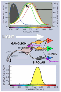 Yellow On – (Blue Off Pathway)
Unlike for red, green, or blue, no cone is most sensitive to a wavelength between the wavelengths that look red or green. Yet yellow is a unique color.
All other colors have appearances that are mixtures of these four. For example, violets and purples look like a mix of red and blue based on whether there is more blue or more red. Oranges mix red and yellow.
While yellow results from mixing red and green light, it has no hint of either. (Similarly, mixing yellow and blue paint pigments results in the absorption of both shorter and longer wavelengths, leaving an intermediate residue of reflected light that looks green without a hint of yellow or blue.)
Yellow comes into the picture by combining the L and M wavelength cone responses. Their combined effect is strongest at their joint midpoint, 555 nm:
The inhibitory bipolar synapse from the S cone pushes the pulse peak to 575 nm for pure yellow.
Blue On – Yellow Off Pathway
You can probably guess. [4]
Still, take a look. It also includes the Y Off response.
Hue is the subjective effect created by the visual system to distinguish differences in wavelength.
 The various color hues are produced by combining the responses in the red, green, yellow, and blue opponent color pathways.
The ability to distinguish differences in hue depends on the combined change in the responses of the three cones as wavelength is changed.
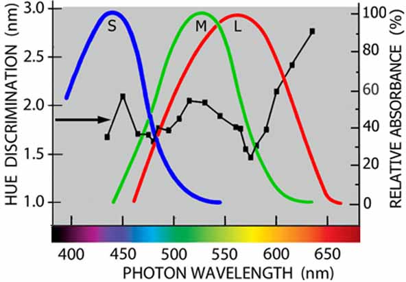
The black line shows our ability to see differences in wavelength based seeing a change in their hue, i.e., wavelength difference-thresholds: [5] (Lower values indicate better discrimination.)
To learn how and why difference-thresholds are measured, see Appendix E – Psychophysical Methods.
Note how the best hue discrimination occurs at 480nm & 575nm, where the overlapping cone functions change most rapidly. (In these regions, small differences in wavelength result in large differences in the cone response ratios.) The average ability to discriminate differences of about 2 nanometers is consistent with our ability to distinguish over 100 different hues.
White On – (Black Off Pathway)
Make the bipolar synapses from all three cones inhibitory, and you have a neural pathway that is released from inhibition by any wavelength -more or less. Its respond is strongest to white light,which has all the wavelengths. Thereby, you get a White On pathway:
Together with inhibition when the cones release glutamate in darkness, the strength of the white response reflects the balance between light and dark.
Black On – White Off Pathway (not shown)
Finally, seeing something that lacks light is as essential for survival as seeing light – like spotting a tick on your skin or reading this text.
Switch the inhibitory and excitatory bipolar-ganglion neural synapses. Now light inhibits the ganglion cell, while darkness releases it from inhibition, resulting in a black pathway.
Besides producing the color effects of black and white to various degrees, the white and black pathways add two additional color effects to the hues produced by the other pathways:
1. the saturation of these hues, such as the difference between red and pink
2. the degree to which the hues are bright or dark, i.e., their value.
Concentric Opponent-Color Pathways
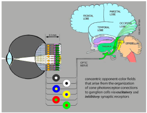
The above figures show how the ganglion neurons are connected via bipolar cells to two or more cones at their location in the retina. Through a lateral geniculate (lgn) synapse their axons connect to corresponding locations in the visual cortex. That is how information in the retinal image is mapped to the cortex. To make the color information in that map more distinct, another network of cone connections is added to the ganglion neurons’ bipolar connection to the cone receptors.
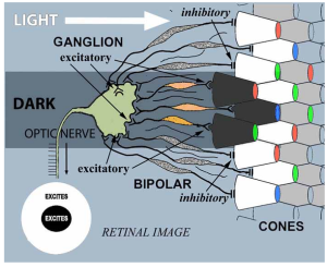
In addition to excitatory and inhibitory connections to their immediate cones, ganglion neurons also have other bipolar connections to cones surrounding their immediate cones. The result is a system of directionally concentric, opponent-color, On-Off pathways (usually called “Opponent-Color Pathways”).
Here a Black Center-White Surround ganglion neuron is excited directly by bipolar neurons that are excited by blue, green and red cones – so it responds to dark at its immediate location. It is also excited by bipolar neurons that are inhibited by surrounding cones. Therefore, it responds most strongly to a black spot surrounded by whiteness.
The other opponent-color pathways also have this center-surround organization added to the direct path shown in the above figures.
The complexity within the ½ mm thin retina is astounding!
 Long before these excitatory-inhibitory pathways were discovered, this opponent-color processing had been proposed by Ewald Herring in 1874. His four color theory was based on three subjective effects:
1. Only 6 colors, red, green, blue, black, white, and yellow are unique. Each has no hint of the other five.
2. All other colors appear to be a mixture of two of these colors.
3. After extended viewing of any one of these colors, there is a tendency to see its opponent color:
Look steadily at the crosshair between the colored squares for 30 seconds. Then, shift your gaze to crosshair between the grey squares.
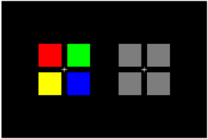 As the pathway for each color depletes its resources, that color’s “on” and “off” effect is removed from the effect of the other pathways. This leaves an opposite color afterimage as the other pathways are stimulated by the light grey.
(Only “sort of” because the adapted color’s “off” effect is also lost.)
To see whether this happens in the visual cortex:
Look at the colored squares with one eye. Then, look at the grey squares with the other.
- Nilsson TH (1972) Effects of pulse duration and pulse rate on hue of monochromatic stimuli, Vision Research,12, 1907-1921. ↵
- Hard A, Sivik L & Tonnquist G (1996) NCS, Natural Color System - from concept to research and applications. Part 1. Color Research and Application, 21, 180-205. Bannert MM & Bartels A (2025) Large-scale color biases in the retinotopic functional architecture are shared across human brains. Journal of Neuroscience, 38, 3657-3668. Tastes are another example of genetically encoded sensations. The five or six types of receptor moleculesin our taste cell membranes bind to specific ionized molecules. Those ions have no characteristics of being salty or sweet any more than photons are red or green. For example, the seemingly universal agreement that sodium chloride tastes salty indicates this subjective effect is genetically determined. ↵
- DeValois RL, Abramov L & Jacobs GH (1966) Analysis of response patterns of LGN cells. Journal of the Optical Society of America, 56, 966-977. Li P, et al (2022) Cone opponent functional domains in primary visual cortex combine signals for color appearance mechanisms. Nature Communications, 13, 6344 {doi.org/10.1038/s41467-022-34020-2}. ↵
- For simplicity, the effects of the inhibitory bipolar connections for the illustrated color ON pathways are omitted in the pulse rate brightness figures at the end of chapter 16 and start of chapter 17. Stimulation by their OFF response wavelengths, inhibits their firing rate. When their ON wavelengths are lacking, stimulation by their OFF wavelengths reduces their firing rate below the random firing rate. This inhibition of firing provides secondary information about the presence of these OFF response wavelengths. Here is a complete pulse-rate response of the Blue On Yellow Off pathway as it varies with the LIGHT’s wave-length: Similar below-random-firing-rate modifications can be added to the other ON-OFF color pathway figures. The wavelength overlap of the S, M, & L absorption functions means that no wavelength can stimulate just the M cones unless the light was focused only on those cones. Fong, et al were able to do this with micro-lasers. The result was a color never seen before. Observersdescribed it as like an intense blue-green. This is consistent with the operation of an internal color palettealigned with wavelength. {Fong J, et al (2025) Novel color via stimulation of individual photoreceptors atpopulation scale. Science Advances, 11, 16;{doi.10.1126/sciadv.adu1052}.
 ↵
- The hue discrimination results are from Nilsson TH (2020) What came out of visual memory: Inferences from difference-thresholds, Attention, Perception & Psychophysics, 82, 2963-2984. ↵