
II. FROM OPTICAL TO VISUAL EFFECTS
16. PHOTON DENSITY AND BRIGHTNESS
As explained in Ch 10, disobeying the Pauli Exclusion Principle enables the density of photons arriving from any specific direction to increase without limit. Thereby, retinal images can differ in photon density over an extensive range. Here are measurements of the effectiveness of photons emitted by various familiar light sources. When corrected in terms of relative visual brightness as a function of wavelength, the energy units change to candela/meter-squared, and the measurement is called luminance. For more information, see Appendix D –Measuring Light.
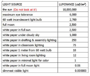
From maximum tolerable to dimmest visible, the visual system operates over a physical intensity range of 2 billion to 1!
That range is amazing, considering that the visual system transmits information about light intensity by neurons, which can only vary their rate of releasing nerve impulses by a factor of about 1 to 25. It responds over this enormous range by using several processes that change its sensitivity (adaptation) depending on how much light is present. These adaptive changes include:
 *1. Most obvious is the change in pupil size between 2 and 8mm diameter. (Appendix F- Directing Your Gaze explains this reflex.) This changes the amount of light entering the eye by a factor of 16X in a few seconds.
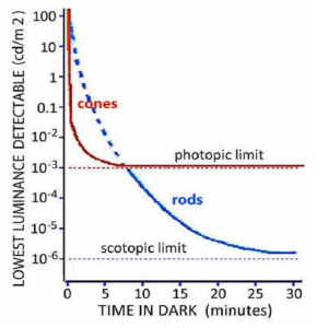
*2. Half of the adaptation involves switching between two systems of light receptors:
2.1 the photopic system, which uses cones for daytime vision
2.2 the scotopic system, which uses rods that are 1000 times more sensitive than cones for vision at dusk and nighttime [1]
*3. Each system can increase its sensitivity 1000-fold in dim light. This dark adaptation can take up to 10 minutes for the cones and over 25 minutes for the rods.
How these systems increase their sensitivity is revealed by their cost.
Visual acuity decreases as the amount of light decreases.
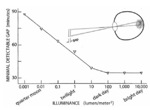
This graph shows how far apart two lines must be to see the gap between them as light decreases: [2]
Over a wide range of daylight, acuity is better than could be expected. A gap scarcely wider than the diameter of a photoreceptor is enough to visually separate the two lines. (Shortly, we’ll learn about several complications that make this acuity remarkable.)
*4. As light decreases, connections between the retinal receptors and ganglion cells change. More retinal receptors connect to each ganglion cell to pool their responses.
This overlap increases the chance that a photon can arrive over a wider range of directions and be captured by one of these receptors. However, each ganglion cell now responds to photons from a wider range of directions. Therefore, the resulting visual response is less sharp.
*5. More neural processing in the retina and cortex can also produce different levels of adaptation in different areas of an image – local adaptation.
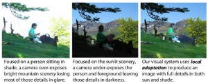 Local adaptation processing can be revealed in certain limited circumstances where it leads to a brightness contrast illusion: despite appearances, the two central squares are equally bright in terms of the amount of light they emit. (Cut two holes in a sheet of paper so you can only see the central squares.)
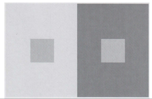
This bit of deception, like many other illusions, is a small price to pay for the enormous benefit of seeing in shadow and sunlight simultaneously.
- A third type of retinal light receptor, melanopsin neurons do not contribute to a visual image. Their photopigments selectively respond to the different levels of daylight over a 24 hour day, with maximal sensitivity at 480nm. Connecting directly to the hypothalamus, their responses drive the circadian sleep and arousal rhythms. See Matin PR (2023) Neurons share an intense load. Science, 379, p.335-336. ↵
- Figure is based on Craik KJW (1939) The effect of adaptation upon visual acuity. British Journal of Psychology, 29, 252-266. ↵