
Main Body
17 ANOTHER PROBLEM WITH PHOTONS:
THEY’RE NOT COLORED! p. 58
Perhaps the most convincing evidence is the changes in hue produced by pulsing single wavelengths of light.17 This indicates that hue perception involves a temporal code which was altered by flashing, as well as by the cone neural pathways.
Let’s consider, “How could the wavelength information available from photons be made evident in a conscious representation?” That picture in our head is filled to the finest possible directional detail available in the retinal image from point to point variation in the number of photons. Adding some kind of symbol to also indicate the wavelength point by point would obscure the directional detail. Alternatively copying the auditory system, by adding some effect like”twinkling” would take time to play out, thereby reducing the speed of perception and ability to notice change.
Somehow evolution hit upon the same kind of solution physicists use when they can’t explain an idea – invent a new “dimension” like strings. Color is an additional dimension in consciousness added to the directional detail of the internal image to represent photon wavelength point by point.
Because photons are not colored, our brain has to do it.
p. 59
The wavelength information gathered by three types of cones is sent to the brain as impulses along six types of neural pathways:

N.B. We are now referring to pathways in the brain where the sensations arise, and not to specific things that might be mistaken as being colored.Therefore, it is OK to use color names. (Sure beats writing “long wavelength pathways” every time – shortening to “long pathways”, etc. doesn’t work : )
These relatively simple neural computations in the retina are the first in series of steps analyzing the relative responses of the three cone receptors.
The next two figures show the basics of how these computations are done.
p. 60
The red excitatory – green inhibitory calculation:
![]()
Note that the LONG wavelength cone absorbs the most photons at 590 nm which looks yellow orange. Pitting the MIDDLE wavelength response against the LONG wavelength response, results in a neural response that peaks at 625, which looks red.
Various non-mammalian animals have three or more types of receptors whose wavelength absorption curves are spread out more evenly. Therefore their systems need less processing to achieve good wavelength discrimination. As primates, we use our brain to make up for poor genetics.
Perhaps that’s how we got started down that road!
Switch the excitatory and inhibitory synaptic connections around, and you get a green excitatory – red inhibitory pathway.
Four to go:
p. 61
Here is a blue excitatory – yellow inhibitory calculation:
It explains how “yellow” comes into the picture. Both the LONG and MIDDLE wavelength  cones inhibit the ganglion cell for this pathway. Note that their combined effect is strongest at the mid point between them, 575 nm, which looks yellow to us.
An excitatory synapse from the SHORT wavelength cone completes the calculation.
To get a yellow excitatory – blue inhibitory – – – . Well, you can probably guess.
Make all the synapses excitatory and you have pathway that responds to any wavelength. Even better if all the wavelengths, white light, are present at once. Thereby one gets a white pathway.
Finally, make all the synapses inhibitory and nothing happens. What good is that? Next page:
Nerves are not inactive when not stimulated. Their sensitivity makes them a bit unstable. Left alone, they irregularly release a nerve impulse, perhaps 1 or 2 every second or so on average. Yet that is also information.
p. 62
The nervous system is often referred to as being “binary” – like current digital computers. But that ain’t so. It is actually a trinary system:
Table 1. Neurons as trinary signal processors.
| impulses per second | neuron’s state | numerical equivalent |
| 0 | inhibited | -1 |
| 1 or 2 random | resting | 0 |
| > 2 | active | +1 |
It can be just as important to know that there is no light coming from a particular direction “out there” as spotting something bright in darkness. Your reading these black letters proves that point.
However, there is more to getting a black pathway.
Some neurons are so “unstable” they fire continuously unless inhibited. Cone activated inhibitory synapses on these result in a true black pathway that actively signals darkness.
Yet there is more. These black and white pathways join onto two other types of ganglion neurons with alternating excitatory and inhibitory synapses to create white excitatory – black inhibitory and black excitatory- white inhibitory pathways. Between them the subtlest differences in shades of gray (i.e. the number of photons) become noticeable.
p. 63
These two pictures illustrate how much information is added by superimposing the information available from photon wavelengths onto the directional detail in a retinal image:18

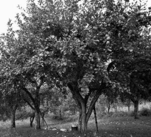
Note how easy it is to see the apples as well as to tell whether they are good to eat.
There is a lot more about color than its hues, but those are other stories.19, 20