
Main Body
12 PHOTONS AS RETINAL IMAGES
p. 37
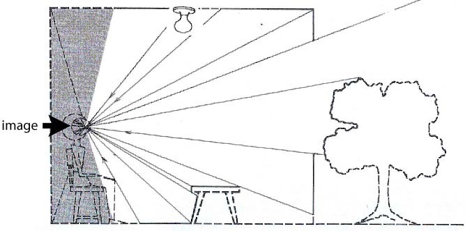
Lenses are how our eyes sort the confusion of rays to produce an image that contains the information about where the rays came from. They do this by forming an image –
p. 38
– an IMAGE like this on the retina at the back of the eye:
When inversion and reversal of the image in our eye was discovered in the Middle Ages, it mystified philosophers.5
The answer to that “little” inconsistency between optics and perception turns out to be more profound than any mystery in physics.
It is not explainable by some possible 1800 twist of neural connections from the retina. Here’s why: After a few days’ confusion, persons wearing “glasses” that turn the image right side up find the world appearing normal.
Evidently the visual image is not constrained by neuroanatomy.
p.39
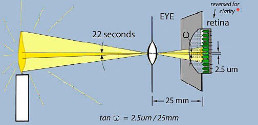
Consider the retinal image. Though it appears to be continuous, that image is sensed by individual receptor cells in the retina.
The ability to sense the direction from which photons have arrived depends on the size of the retinal receptors and focal length of the eye. A typical photoreceptor has a diameter of 2.5 micrometers and is located at the focal point of the eye, some 25 millimeters behind the lens. By trigonometry the receptor subtends a 22 second angle with respect to the lens’ center. Because congruent angles are equal, that is the receptor’s field of view – how much of the world it responds to.
*NB In vertebrate eyes the light sensitive end of the photoreceptors not only faces away from the lens, it is also covered by the rest of its cell body and by several types of nerve cells and nerve fibers that, fortunately, are nearly transparent. (Why this backwards design, and only in vertebrates? That’s another story.)6
p. 40
However, the photoreceptors would not respond to the photons in the image were it not for another property of photons: